June 2026
The full team is together and data collection begins!
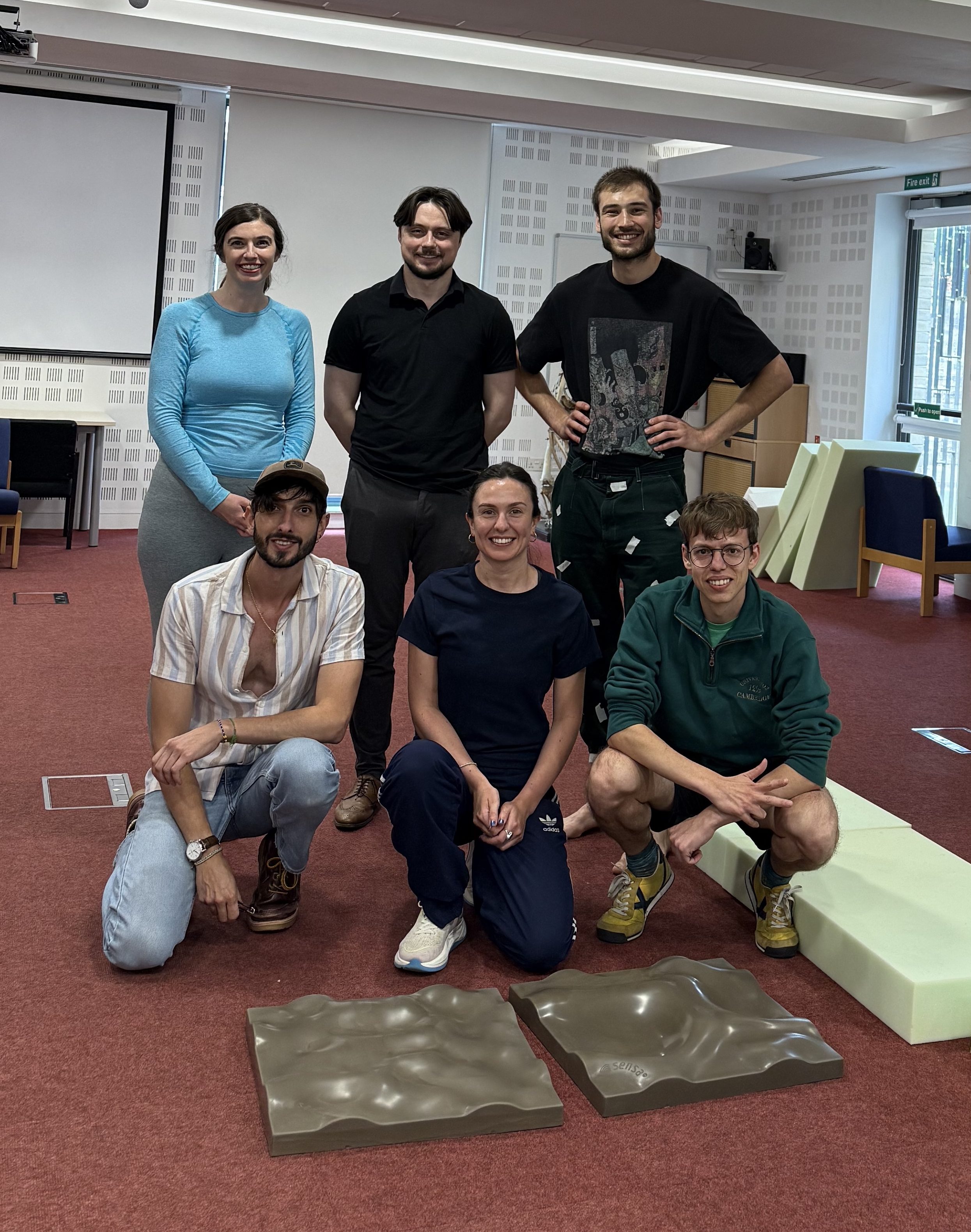
From top left to bottom right: Ashleigh Wiseman (PI), Paul Minhoff (Cambridge PhD student), Anxo Rubalcaba Herrero (CISPAC PhD student), Grégoire Boulinguez-Ambroise (STEPS Researcher), Ella Lipscome (Project Coordinator), and Oriol Monclús-Gonzalo (STEPS Researcher).
June 2026
Welcome to the team, Dr Grégoire Boulinguez-Ambroise!
Dr Grégoire Boulinguez-Ambroise joins the STEPS Team as our new Postdoctoral Researcher. He will be based at the McDonald Institute for Archaeological Research in Cambridge with Dr Wiseman.
In the context of the STEPS project, Dr Boulinguez-Ambroise’s role focuses on the collection and processing of experimental biomechanical data, alongside investigating how the human body adapts to different types of movements.
April 2026
New paper published!
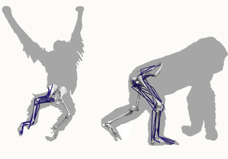
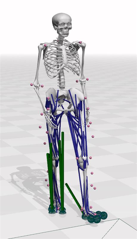
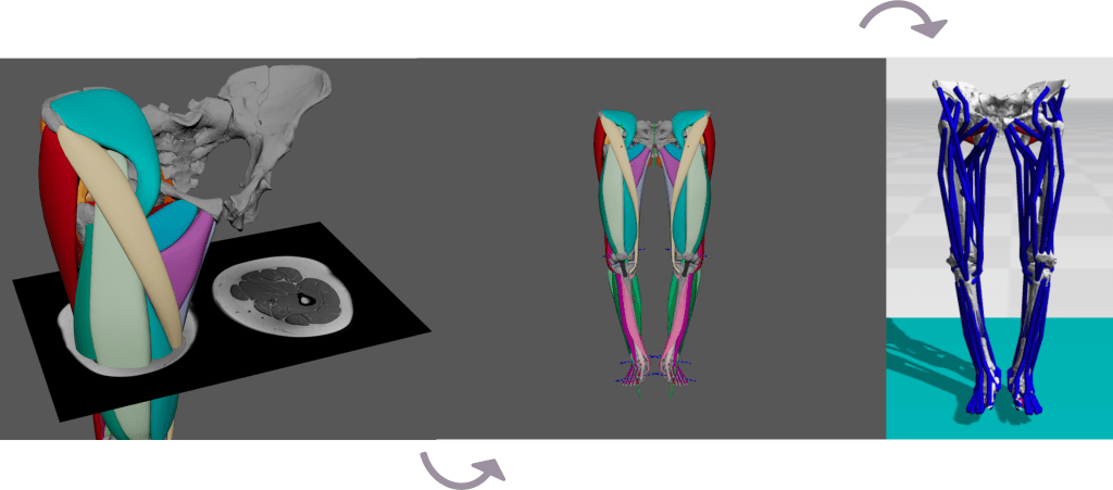
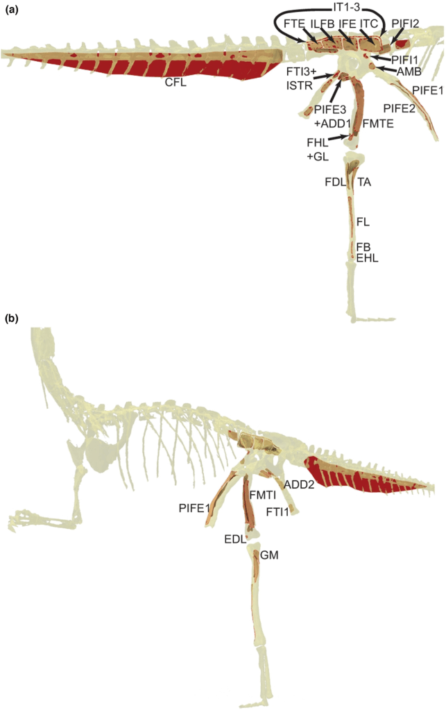
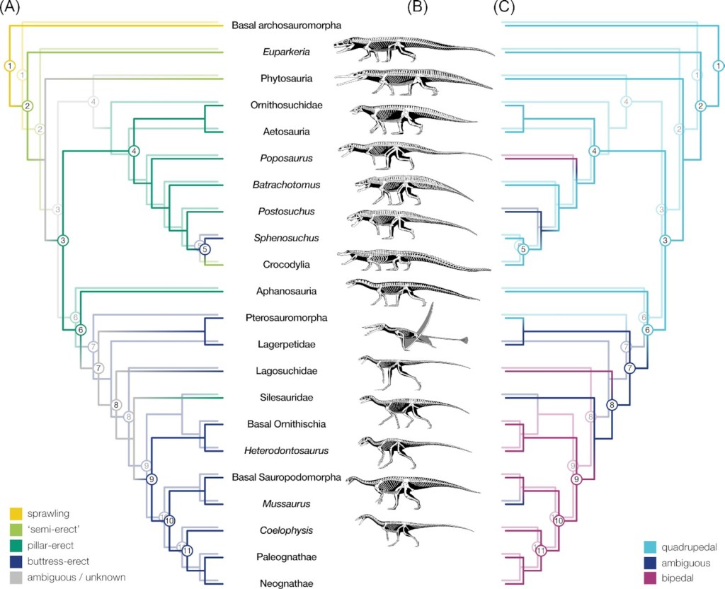
A new study, published in Royal Society Open Science, found that muscle function differs systematically between primate species and reflects their typical modes of movement.
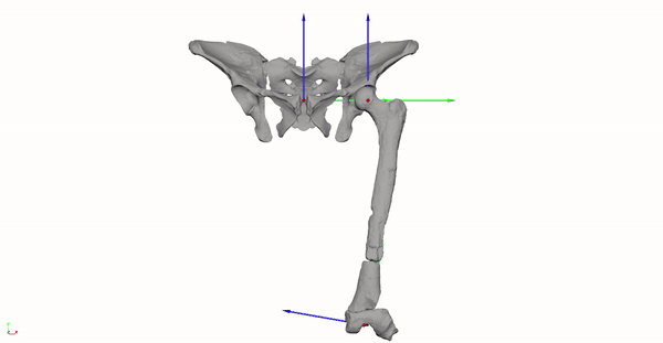
The study investigates how differences in limb anatomy shape the way primates move. By building detailed, subject-specific 3D musculoskeletal models of the hind limbs in a range of primate species the researchers measured how effectively muscles generate movement at different joint positions.
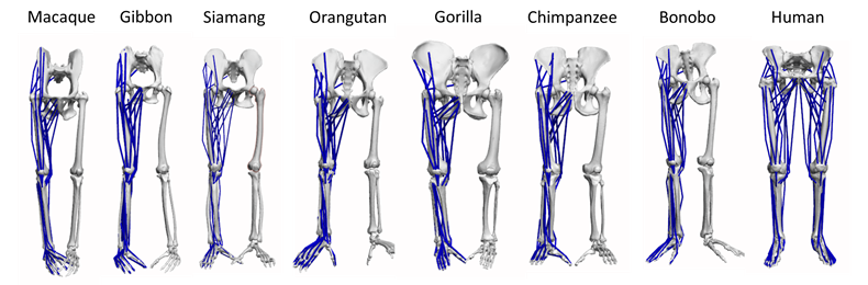
These measurements, known as moment arms, provide insight into the mechanical advantage of muscles and how this varies across species with different locomotor behaviours.
In highly arboreal primates such as orangutans, gibbons, and siamangs, muscles around the hip show large and flexible ranges of mechanical advantage, supporting control across a wide variety of postures needed for climbing and moving through trees.
In contrast, humans show peak muscle effectiveness in more extended, upright positions, consistent with the demands of bipedal walking and running.
Importantly, these differences are not simply a matter of size, but reflect shifts in when muscles are most effective during habitual movement. This work provides a new comparative framework for understanding how anatomy, movement, and evolution are linked across primates.
Written by Lydia Clough & Ashleigh Wiseman
April 2026
Welcome to the team, Dr Oriol Monclús-Gonzalo!
Dr Monclús-Gonzalo joins the STEPS Team as our new Postdoctoral Researcher. He will be based at the McDonald Institute for Archaeological Research in Cambridge with Dr Wiseman.
In the context of the STEPS project, Dr Monclús-Gonzalo’s role focuses on the collection and processing of fossil and comparative 3D data, the cataloguing of postcranial variation, and contributing to the creation of musculoskeletal models, including the development of predictive models to estimate muscle architecture parameters from osteological correlates.
March 2026
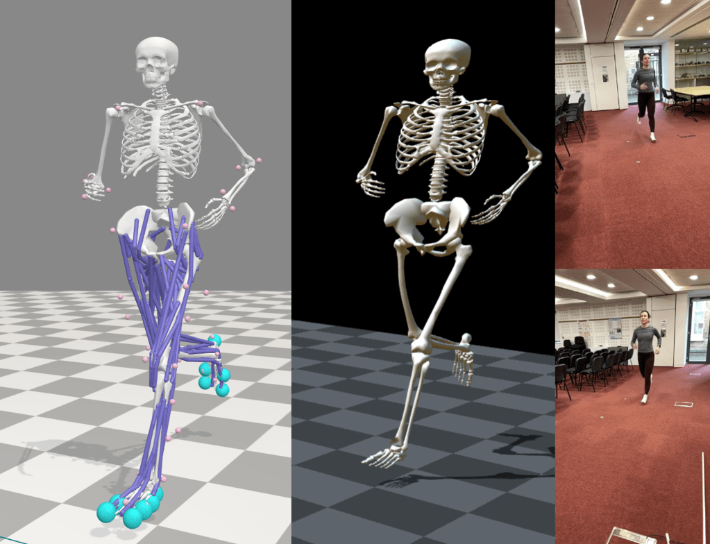
Preliminary experiments begin
The team started testing OpenCap to determine its usefulness in our planned data collection for the STEPS project. We tested different camera setups, positions, and track lengths, to varying degrees of success (note: we struggled to get consistent kinematics across the different camera configurations!). If anyone is interested in reading our technical report on this, reach out at steps@arch.cam.ac.uk.
February 2026
The team expands
Ella Lipscombe joins the STEPS Team as our new Project Coordinator. Welcome to the team, Ella! Ella will be based at the McDonald Institute for Archaeological Research in Cambridge with Dr Wiseman.
September 2025
ERC Starting Grant awarded
Dr Wiseman was awarded a five year grant by the ERC for the project STEPS (2025-2030). Read about it here. The project will start on November 1st 2025 and will support the employment of four people: the PI, two PDRAs and one Project Coordinator. We will be supported by external collaborators across the world.
January 2025
New publication! Read it here.
Abstract: “Endurance running is thought as critical for the evolutionary success of hominins. A new study analysing the running skills of the famous ‘Lucy’ – Australopithecus afarensis – finds that they performed poorer than modern humans, suggesting that key features of the human body plan evolved specifically to improve running performance.” – Wiseman, 2025.
December 2024
Data collection at the Jan Palfijn Lab in Belgium
Dr Wiseman travelled to Belgium to study the musculoskeletal anatomy of a gorilla and an orangutan with Dr Julia van Beesel and Professor Evie Vereecke at the KU Leuven! We were also joined by Pasha van Bilijert. We collected some Diffusion Tensor Imaging data at UGent with the assistance of Océane Cluzeau.
September 2024
Workshop in Barcelona, Spain
Dr Wiseman and Dr Demuth travelled to Barcelona to attend a workshop on PredSim and biomechanical simulations, organised by staff at the KU Leuven.
August 2024
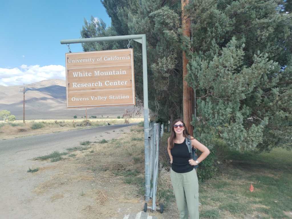
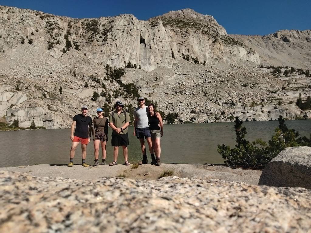
Workshop at the White Mountain Research Center, California
Dr Wiseman, Dr Julia van Beesel and Dr Oliver Demuth travelled to the White Mountain Research Center to attend a workhop on Phylogenetic Comparative Methods for Biomechanists, hosted by Dr Daniel Moen from UC Riverside.
Funded by the National Science Foundation grant (IOS-2430681), awarded to Daniel Moen.
July 2024
Data collection at the Jan Palfijn Lab in Belgium
Dr Wiseman travelled to Belgium to study the musculoskeletal anatomy of a macaque and a siamang with Dr Julia van Beesel and Professor Evie Vereecke at the KU Leuven! We collected some Diffusion Tensor Imaging data at UGent.
March 2024
AABA Cobb Professional Development Grant awarded!
Dr Wiseman was awarded an AABA Cobb Professional Development Grant for the project Beyond the bones: Enhanced phylogenetic bracketing of hominin soft tissues. This project will conduct primate dissections and explore diffusion tensor imaging of primate musculature! This work will be conducted in collaboration with Professor Evie Vereecke and Dr Julia van Beesel at the Jan Palfijn Lab, KU Leuven. The first data collection trip is planned for summer 2024.

January 2024
New publication!
How reliable is musculoskeletal modelling in producing replicable outputs? A study aiming to understand how different methods of estimating muscle structure influence our interpretations of limb function
Read the blog post by Ashleigh L Wiseman and Lydia Clough: https://www.arch.cam.ac.uk/news/Lucy-modelling
November – December 2023
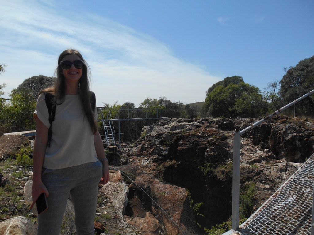
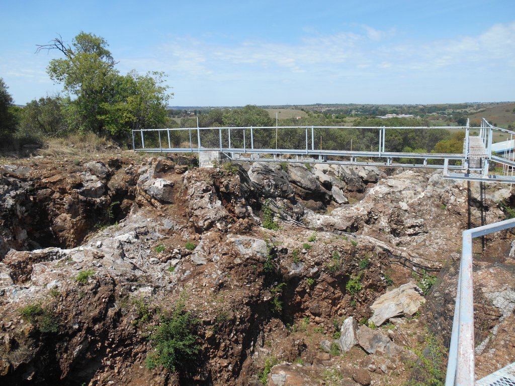
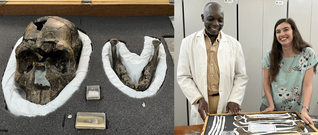
Research trip to South Africa
In late November 2023, Dr Wiseman travelled with colleagues from the the UK and France to Johannesburg, South Africa.
They visited the hominin fossil collections at Wits University, toured the Cradle of Humankind and the Sterkfontein cave system with Dr Dominic Straford, and attended a two day long workshop called BrAIn, organised by Dr Amelie Beaudet.
September 2023
Data collection at the Jan Palfijn Lab in Belgium
Dr Wiseman travelled to Belgium to study the musculoskeletal anatomy of a chimpanzee and a gibbon with Dr Julia van Beesel and Professor Evie Vereecke at the KU Leuven! We were assisted by two wonderful interns: Simon Debusschere and Arthur Arie.
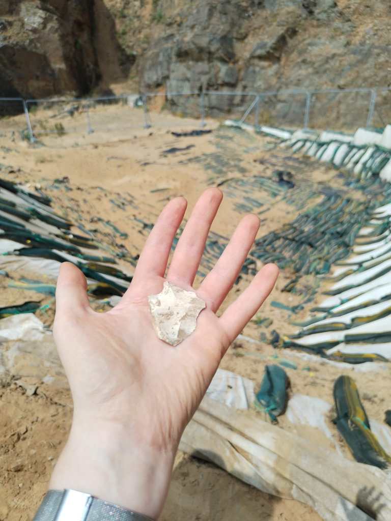
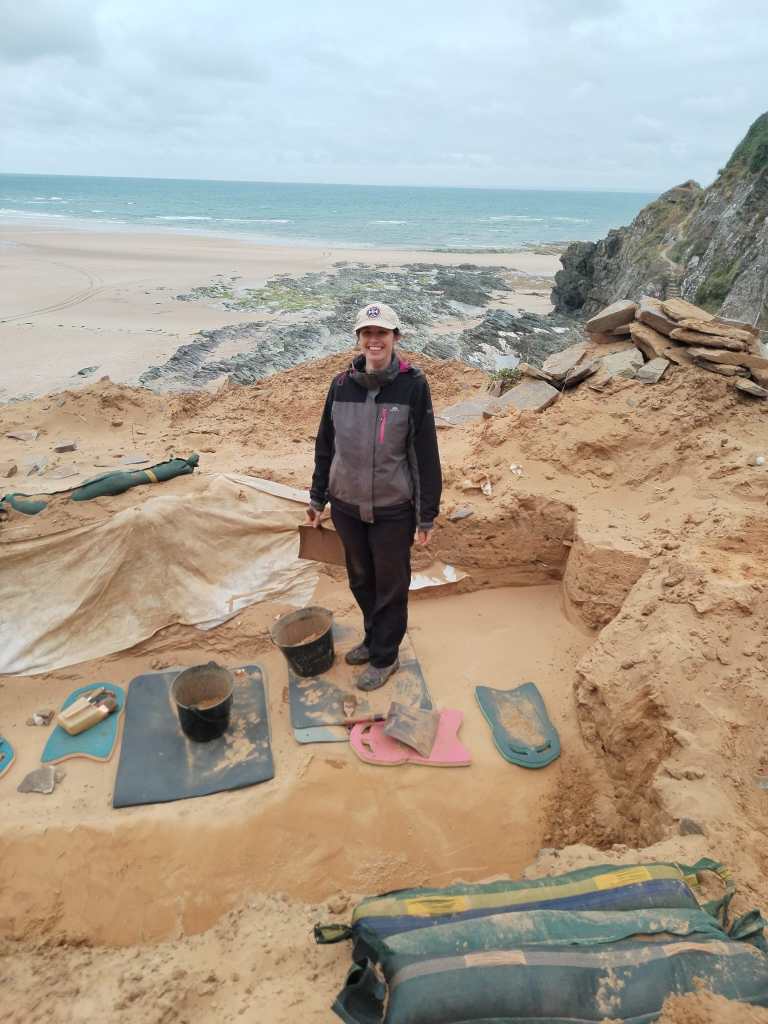
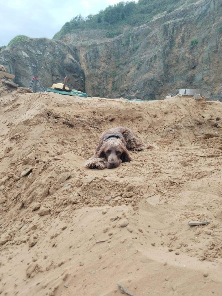
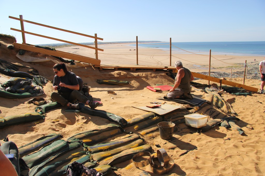
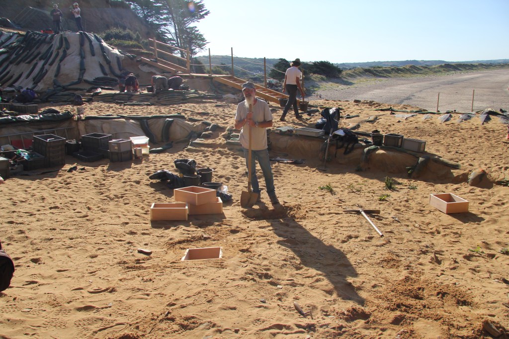
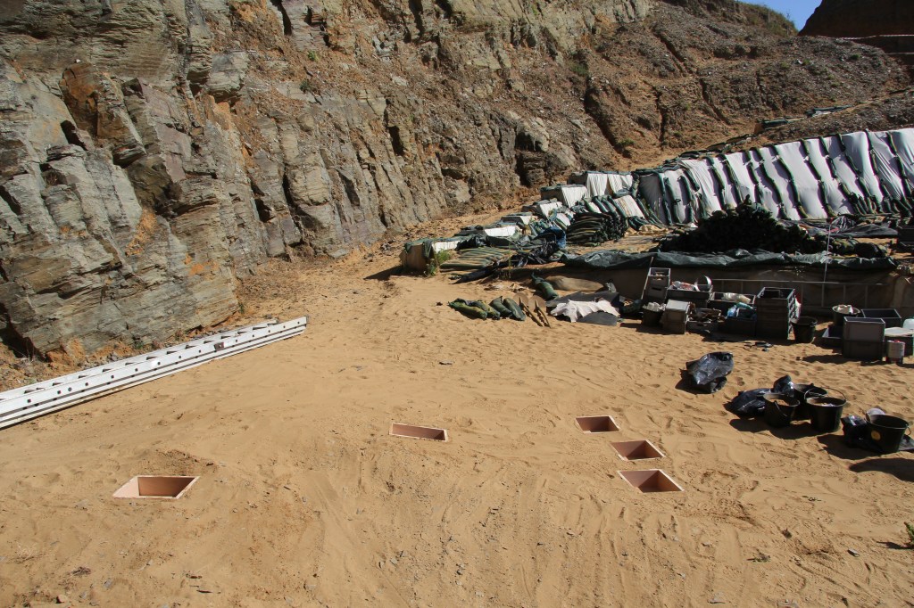
June-July 2023
Fieldwork at Le Rozel, France
Dr Wiseman returned to Le Rozel for futher fieldwork at the rock-shelter site. It was a productive season! And it was made even better by the addition of the new site doggo!
This work was funded by a McDonald Fieldwork and Travel Grant, University of Cambridge.
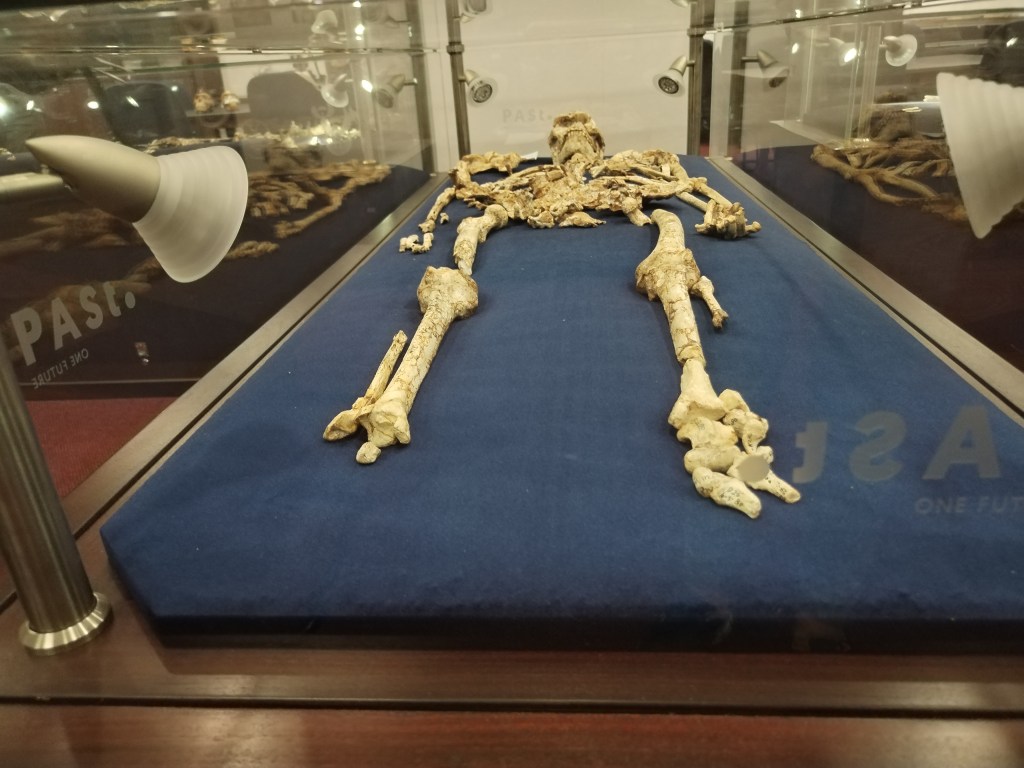
June 2023
New publication!
Dr Wiseman has digitally reconstructed the missing soft tissue of an early human ancestor – or hominin – for the first time, revealing a capability to stand as erect as we do today.
Dr Wiseman has 3D-modelled the leg and pelvis muscles of the hominin Australopithecus afarensis using scans of ‘Lucy’: the famous fossil specimen discovered in Ethiopia in the mid-1970s.
Press release here.
March 2023
Two new publications!
1. Quantitative biomechanical assessment of locomotor capabilities of the stem archosaur Euparkeria capensis
2. Modern three-dimensional digital methods for studying locomotor biomechanics in tetrapods
References:
Demuth OE, Herbst E, Polet DT, Wiseman ALA, Hutchinson JR. 2023. Modern three-dimensional digital methods for studying locomotor biomechanics in tetrapods. J Exp Biol. 226(Suppl_1).
Demuth OE, Wiseman, AL, Hutchinson JR. 2023. Quantitative biomechanical assessment of locomotor capabilities of the stem archosaur Euparkeria capensis. R Soc Open Sci. 10 (1): 221195. https://doi.org/10.1098/rsos.221195

January 2023
Research trip to Nairobi, Kenya
Data collection was funded by Dr Wiseman’s Leverhulme Trust Early Career Fellowship (grant no. ECF-2021-054), the Isaac Newton Trust (Project_21.08(a)), and further supported by the Ng’ipalajem Project (ERC, Grant No. 101020478), awarded to Professor Marta Mirazon Lahr.
December 2022
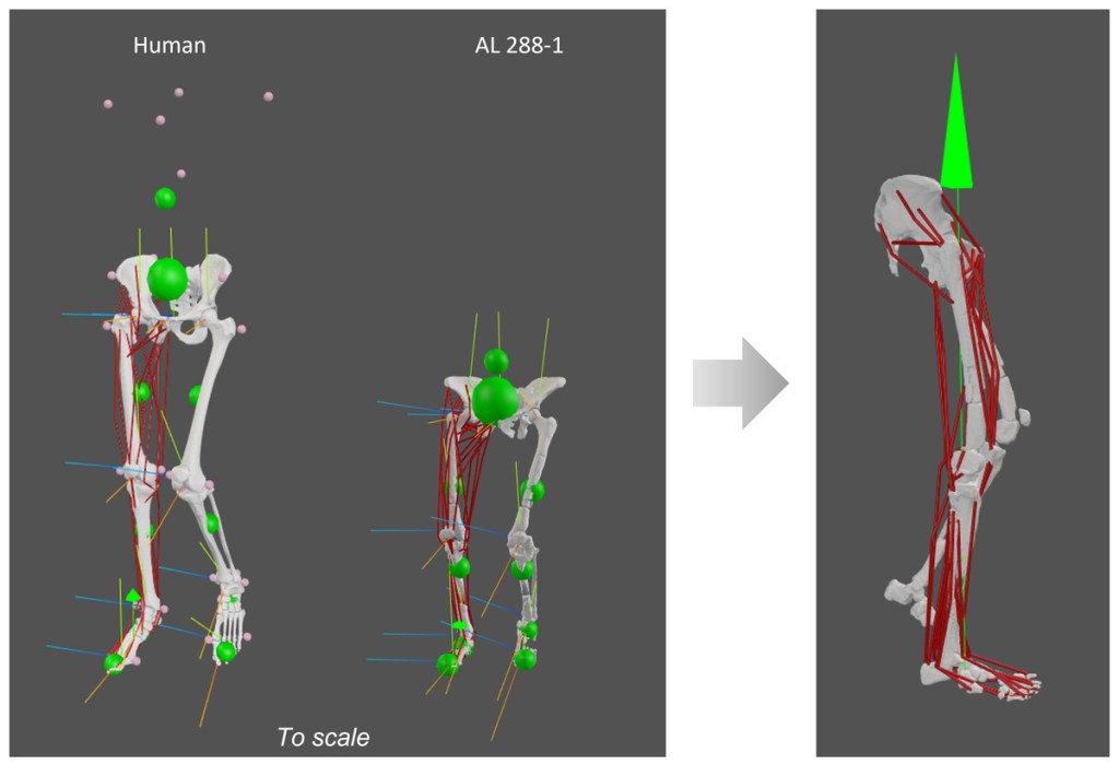
Preprint now available on BioRxiv which details a 3D polygonal muscle modelling approach in the Australopithecus afarensis specimen AL 288-1! See the blog post here about the paper, which also includes videos of the muscles from superficial to deep layers!
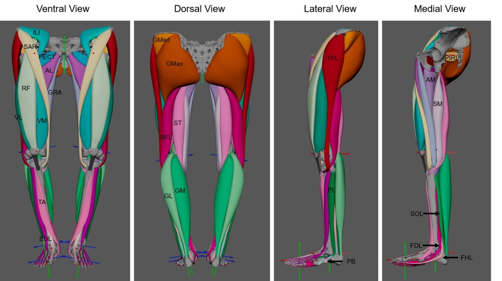
So, why do we need to use such an approach? Musculature simply does not survive in the fossil record. Rather, we are left looking at just the bones. But muscles animate movement, allowing an organism to walk, run, jump and even dance. So if we want to understand how an extinct specimen might have moved, then we need to understand its musculature. Typically, palaeontologists/palaeoanthropologists use a method called the Extant Phylogenetic Bracket to accomplish this task which involves identifying the closest living analogy to the extinct specimen to estimate where muscles attached and how they lay in the body. In the past, this produced simple straight lines of action from a point of origin to the insertion location, which fails to capture how the muscle might have wrapped around bones. In the last 20 years, wrapping surfaces and via points have been developed in biomechanical modelling software to target this problem. The 3D polygonal modelling approach expands upon this problem even further by considering how much space a muscle might have occupied and its path within the body (i.e., this muscle lies on top of this muscle and extends in this direction, so forth). To accomplish this, scaled MRI cross sectional scans were used to define muscle boundaries, allowing the creation of 3D muscles which occupy a realistic space within the body.
This is a necessary precursor to future dynamic research. This 3D modelling approach shows great promise for recreating the musculature of other hominins… stay tuned!
November – December 2022
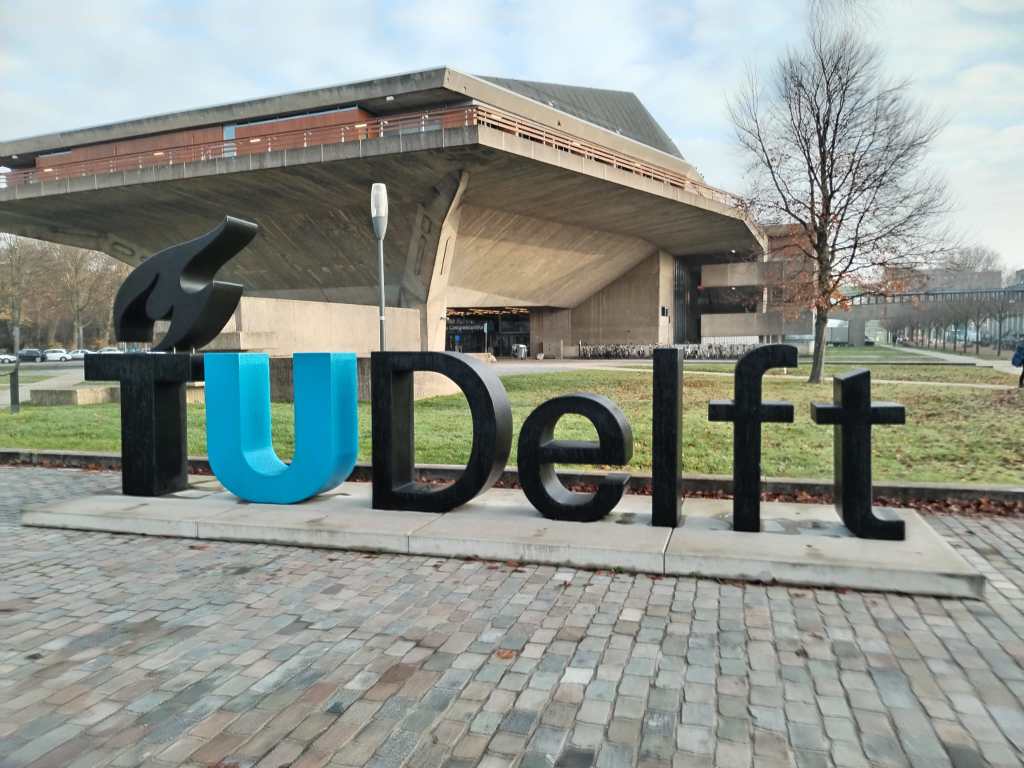
Research trip to the TU Delft
Dr Wiseman travelled to the Computational Biomechanics Lab at the TU Delft, Netherlands for a few weeks to chat research, dynamics and simulations.
August 2022
New publication!
How to reconstruct the muscles of fossil specimens from the attachment sites on bones? Read it here.
July – August 2022
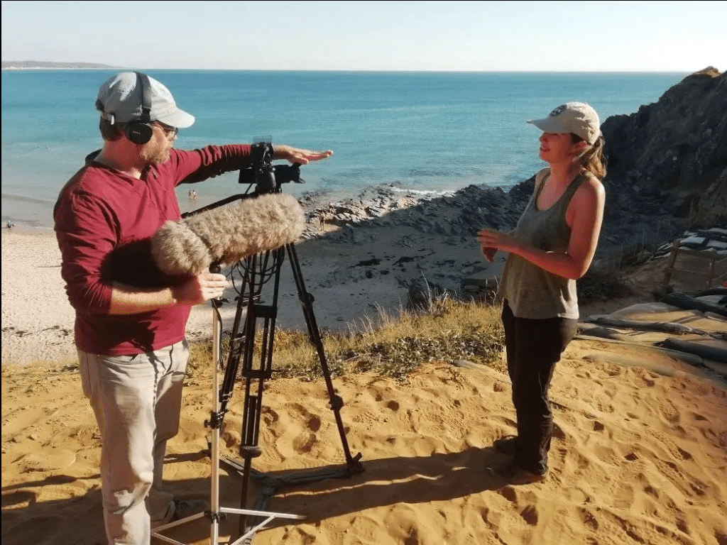
Fieldwork and documentary filming at Le Rozel, France
In addition to fieldwork and experiments, Dr Wiseman was involved with Néandertal – Dans les pas d’une autre humanité, a documentary produced by Court-Jus Production. Read more about it here.
The documentary filmed our excavations and experiments at the site, and even managed to capture new fossil footprint discoveries as they happened.
In the documentary, researchers and scientists debated what life would have been like for the Neanderthals 80,000 years ago, and how the site of Le Rozel fits in with other sites across Northern Europe.
The documentary was released in Spring 2023 and was featured in a number of film festivals across Europe.
In October 2023, the documentary was translated into English and released titled “Neanderthal, in the footsteps of another Humanity”. The documentary can be viewed online.
This work was funded by a McDonald Fieldwork and Travel Grant, University of Cambridge.
August 2022
Paper published!
Reconstructing articular cartilage in the Australopithecus afarensis hip joint and the need for modelling six degrees of freedom
Read it here!
What is it about? It is unusual for soft tissues to preserve in the fossil record and, therefore, cartilage is very poorly preserved. In the case of the famous Australopithecus afarensis AL 288-1 skeleton (nicknamed ‘Lucy’ after the excavators found her whilst listening to Lucy in the sky with diamonds by The Beatles – read more about the discovery here), around 40% of her skeleton has amazingly been found – this is one of the most complete hominin skeletons discovered to date. But only the hard tissues have preserved – that is, the bones. In each of the joints of a body, articular cartilage is preserved which helps with the dissipation of stress during joint movement. Cartilage can be thick or thinly distributed through the joint’s articular surface (or a mixture of both) depending on the joint and its functionality. So if no cartilage has preserved belonging to Lucy, how can we accurately rearticulate her skeleton? And if we under- or over-predict the joint spacing, what repercussions could this have for claims regarding movement?
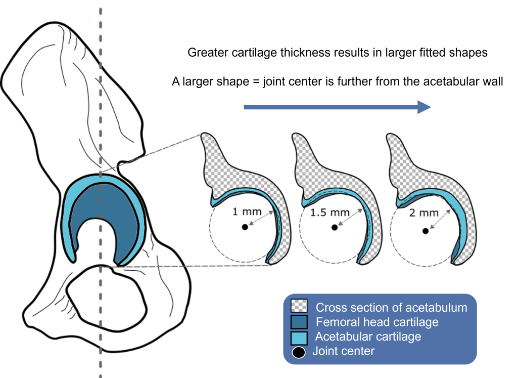
ROM mapping methods have been developed in recent years for a select range of extinct and extant species to ascertain how two body segments articulate and move relative to one another. ROM mapping relies upon movement of a body segment around a joint center and can encompass rotational and/or translational movement. The method identifies which poses are viable and which are non-viable based on bone morphology, thus providing information regarding limb posture.
We employed ROM mapping methods to estimate the joint spacing of AL 288-1’s hip joint in comparison to a modern human and chimpanzee. Nine simulations assessed different joint spacing and tested the range of joint congruency (i.e., ranging from a closely packed socket to loosely packed). We further evaluated the sphericity of the femoral head and whether three rotational degrees of freedom (DOFs) sufficiently captures the full ROM or if translational DOFs must be included.
We tested the articulation and possible osteological ROM of the AL 288-1 hip joint by modelling a static single axis translation to investigate increasing joint spacing, which was considered a proxy for measuring maximum cartilage thickness. We expanded upon this by including all six DOFs, thereby reflecting true joint movement. Whilst the resultant ROM maps were quite similar, there was a greater spectrum of viability in the six DOF simulation than the other simulations, in which the femur was capable of osteologically moving into a greater range of poses. With this spectrum of poses, AL 288-1 was capable of a repertoire of movements, such as erect bipedalism across a range of substrates at various speeds and vertical climbing. Overall, six DOFs are a requirement for modelling mobility in fossil hominins, otherwise the resultant functionality of a given joint may be wrong.
We conclude that the likely maximum joint spacing/cartilage thickness of AL 288-1’s hip joint was 2.448 mm which is on par with allometric scaling assumptions (i.e., the smaller bodied AL 288-1 has a more cartilaginous hip joint than the larger bodied human and chimpanzee). Similar estimates were also generated from the single axis translational simulations, despite some implied functional limitations.
The important bit: we cannot ignore translational movement of the hip when estimating the motion-capability of extinct species!
May 2022
New paper published!
‘Walking—and Running and Jumping—with Dinosaurs and their Cousins, Viewed Through the Lens of Evolutionary Biomechanics’, By Cuff et al, 2022. Read about it here.
March 2022
New paper published!
How to reconstruct musculature in extinct species using digital tools. Read about it here
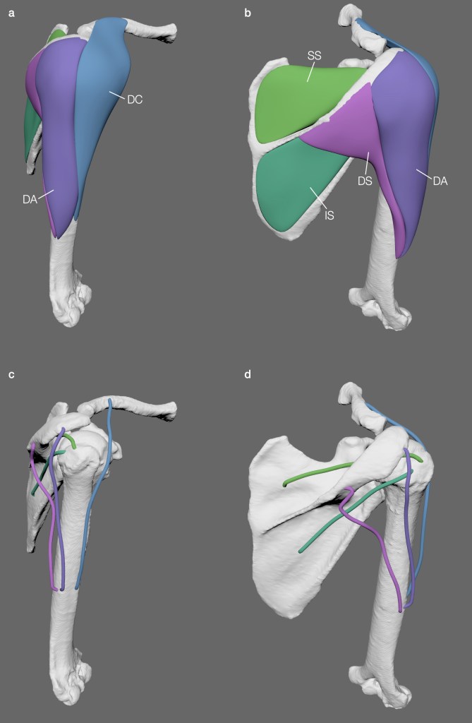

Modelling muscles in a gorilla shoulder (left) and in Euparkeria capensis (right)
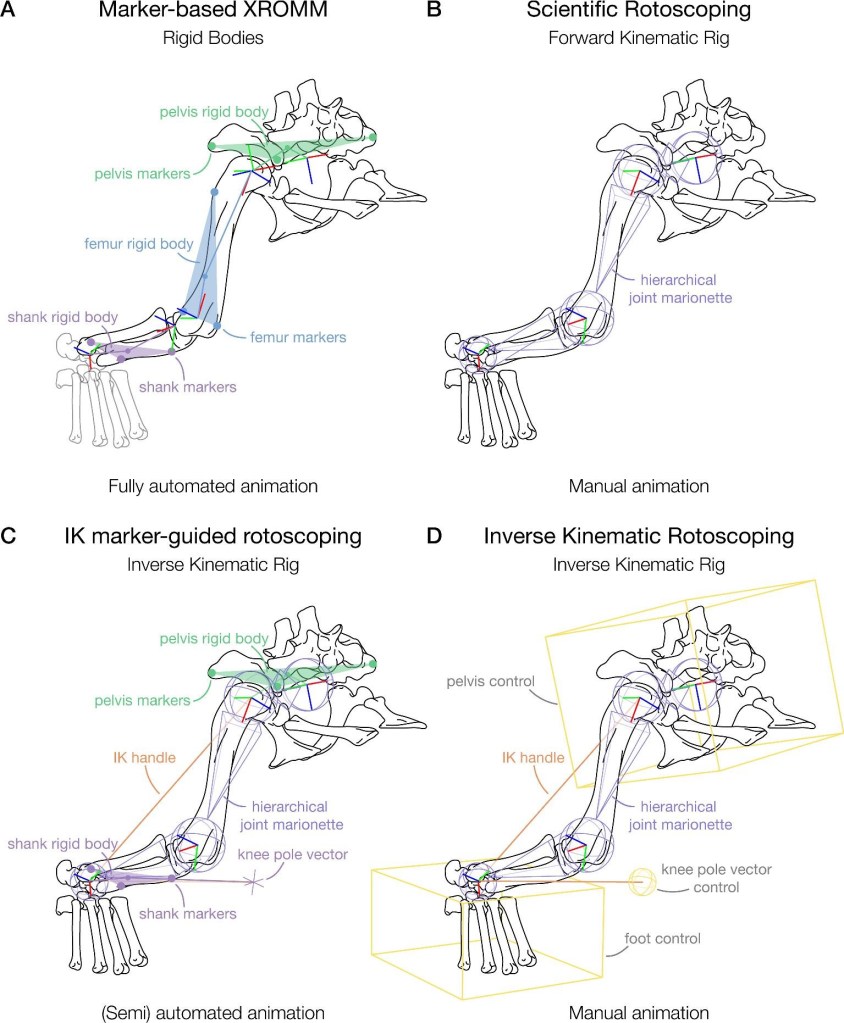
January 2022
New paper published!
IK rotoscoping procedures in XROMM when you have too few markers or markers go missing?
Read about the paper here.
January 2022
Two new publications!
Fossil footprints at Formby Point, UK.
Wiseman*, A.L.A., Vicari, D., Belvedere, M., De Groote, I. 2022. Neolithic track sites from Formby Point, England: New data and insights. Journal of Archaeological Science: Reports, 44, 103546.
Wiseman*, A. L. A., De Groote, I. 2021. One size fits all? Stature estimation from footprints and the effect of substrate and speed on footprint creation. The Anatomical Record 1-9.
October 2021
Leverhulme Trust Early Career Fellowship begins
Dr Wiseman was awarded a Leverhulme Trust Early Career Fellowship alongside an Isaac Newton Trust Fellowship (University of Cambridge) to explore the musculoskeletal anatomy of hominin species. The project will run from 2021-2024 and will be based at the McDonald Institute for Archaeological Research, University of Cambridge